
GenesisScope®Real-time Embryo Monitoring System
A time-lapse imaging and analysis platform designed for embryology research and livestock breeding applications. Within a stable culture environment, the system continuously captures high-definition time-lapse images of multiple embryos with minimal disturbance, recording the complete developmental process from early stages to blastocyst. It provides researchers with objective and traceable morphological and morphokinetic data, and leverages AI algorithms to support studies of embryonic development, culture optimization, and data-driven breeding decisions.
24/7
Continuous real‑time monitoring
0
Disturbance to culture conditions
HD
High‑definition image records
AI
Intelligent analysis algorithms
A Complete Solution for Embryo Development Analysis
Through the deep integration of precision imaging devices, an intelligent controller, professional analysis software, and a customized culture dish, GenesisScope® forms an end‑to‑end solution for embryo development observation and data analysis, serving both scientific research and livestock reproduction.
Imaging Unit
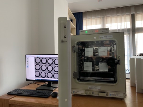
Installed directly on the internal shelf of the incubator. Place the culture dish with embryos in the field of view to start non‑disruptive, continuous monitoring of the entire development.
Provides continuous, high‑definition imaging of embryos in a stable culture environment.
Control Computer
Located outside the incubator and connected via dedicated data cables, fully isolating electronic components from the culture environment.
One controller centrally manages up to six imaging units, improving laboratory throughput and efficiency.
Analysis Software
Supports continuous observation and image acquisition throughout embryo development, enabling both morphological and morphokinetic annotation for comprehensive scientific analysis.
Image generation, parameter configuration, data storage, and intelligent analysis.
Multi‑well Embryo Culture Dish
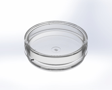
Custom‑designed for the system, featuring 12 wells with coordinate markings to ensure precise embryo tracking.
Provides a safe micro‑environment for embryo culture and enables accurate positional registration within the system.
Imaging Unit
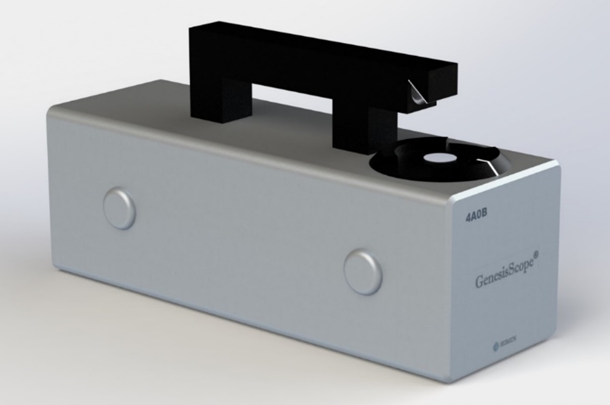
Innovative Optics to Reveal Developmental Details
The microscope implements an innovative Hoffman modulation contrast imaging principle, enabling clear, relief‑like visualization of transparent embryo cells so that key details such as nuclei, polar bodies, and cell division events are clearly rendered.
Hoffman Modulation Contrast
Delivers clear, three‑dimensional relief‑like images that reveal nuclei, polar bodies, cleavage events, and other critical details.
Ultra‑high Resolution
5‑megapixel industrial camera with 2.2×2.2 μm pixel size and 0.6 μm optical resolution.
Embryo‑friendly Illumination
Pure 550 nm green light minimizes phototoxicity and helps protect embryo viability.
Compact design and precise focusing
24×8×12.2 cm
Extremely Compact
Maximizes the available space inside the incubator.
Saturated humidity
Sealed Design
Engineered for long‑term stability even under demanding conditions.
±0.2 μm
Precise Focusing
Ensures repeatable positioning accuracy for multi‑layer scanning.
Ultra‑low Power and Gentle Operation
Industrial‑grade control and imaging electronics combined with optimized algorithms significantly reduce operating current, thereby minimizing heat generation and electromagnetic interference at the source.
5 V
Operating voltage
<20 ms
Exposure time
<10 s
Cumulative light dose
Control Computer
As the intelligent hub of the system, the controller orchestrates the operation and data acquisition of multiple imaging units, turning complex multi‑device time‑lapse experiments into a stable and efficient integrated workflow.
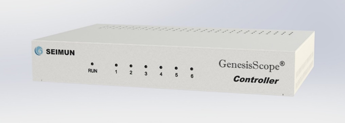
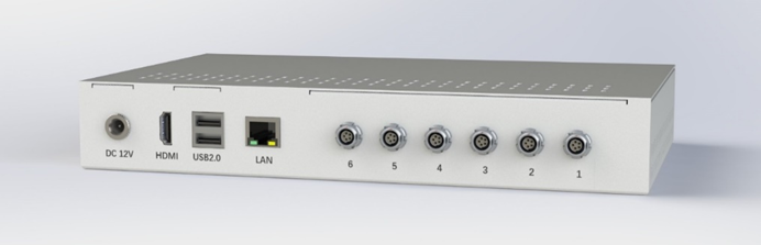
Industrial‑grade Computing Platform
Standard 1U rack‑mount industrial PC with Windows 10, 8 GB memory, and a 120/240 GB SSD, providing robust performance for smooth operation and large‑scale data processing.
Visual Status Monitoring
Front‑panel status indicators clearly reflect the connection and operating status of each imaging unit, allowing researchers to quickly understand system health and enhance safety and manageability.
Highly Expandable I/O Design
Rear panel integrates dedicated ports for up to six imaging units, plus HDMI, USB, and RJ45 network interfaces for display output, data exchange, and network access, enabling seamless integration into existing lab environments.
Monitoring Software
Brings device management, parameter configuration, and time‑lapse project control together in one intuitive interface — the intelligent command center of the system.
Device Management
Centrally manages all connected imaging units.
Parameter Settings
Provides flexible control over key imaging parameters.
Project Control
Accurately controls time‑lapse tasks for each imaging unit.
A Human‑centric, Seamless Workflow
Auto‑detection and Initialization
Plug and play
The controller automatically detects all connected imaging units, identifies device IDs and firmware versions, and completes initialization so devices are ready to use in minutes.
Real‑time Tuning and Optimization
What you see is what you get
Enter live mode from the device list to adjust focus, brightness, or test multi‑layer scans. All changes are displayed in real time so you can quickly optimize image quality.
One‑click Start and Full Monitoring
Intelligent tracking
Start time‑lapse projects with a single click, monitor project status in real time, review the latest images and multi‑layer scan results, and follow the entire embryo development timeline.
Flexible Project Management
Mid‑course intervention
Powerful mid‑project control enables pausing, medium change, and other interventions, improving both experimental flexibility and success rates.
Embryologist
A professional analysis tool tailored for embryology laboratories, turning complex embryo assessment workflows into structured, data‑driven processes to support objective analysis and informed decision‑making.
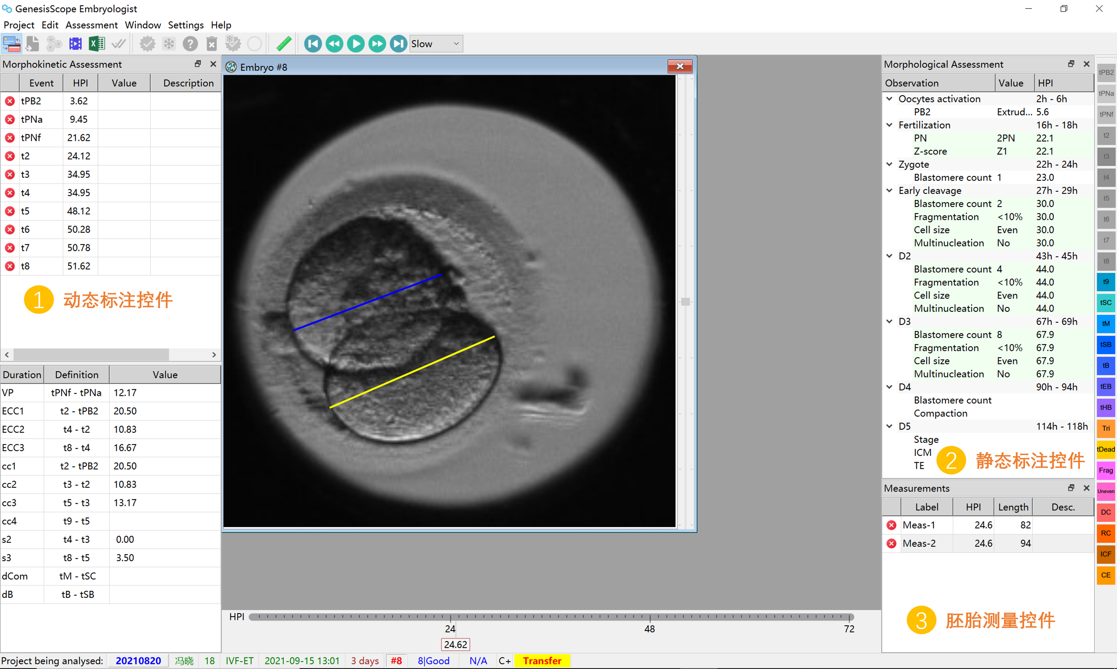
In‑depth Single‑embryo Analysis
Microscopic insight
Freely zoom into images to inspect cellular‑level details, use measurement tools to obtain precise morphological metrics, and add annotations to document key findings.
Global Synchronized Comparison
Macroscopic perspective
Displays all embryos in a well‑organized matrix and plays them in sync frame by frame, making it easy to compare developmental progress and identify the most promising candidates.
Intelligent Annotation and Analysis
AI‑powered
Manual mode allows precise time‑point annotations, while AI mode automatically labels key events and computes morphological and morphokinetic parameters.
Custom Smart Ranking
Quantitative evaluation
Define scoring rules based on clinical experience. The system automatically calculates scores for all embryos and performs smart ranking and categorization.
One‑stop Data Management
Efficient and convenient
Supports statistics, raw data export, and one‑click generation of standardized embryo evaluation reports.
Outstanding Compatibility
Seamless integration
Supports importing time‑lapse projects from Primo Vision dishes with 9‑well or 16‑well layouts.
Embryo Culture Dish (12 Wells)
Premium MaterialSafe and reliable
Manufactured from USP‑compliant polystyrene to provide a biologically safe culture environment for embryos.
Independent Micro‑wellsAccurate localization
The base is laid out as a 3×4 matrix, forming 12 independent wells. A coordinate system based on rows (1, 2, 3) and columns (A, B, C, D) enables precise embryo identification and traceability.
Compact Form FactorEasy handling
With a maximum diameter of just 38 mm, the dish is highly compact and easy to manipulate and store in daily lab workflows.
38 mm
Maximum diameter
12
Independent wells
3×4
Matrix layout
Safety Certifications and Test Standards
Strictly follows international standards to ensure the safety and reliability of every batch of culture dishes.
Sterile Processing
Sterilization process compliant with ISO 11137.
SAL 10⁻⁶
Sterility assurance level
Non‑cytotoxic
Validated to be free of cytotoxic effects.
USP <87>
ISO 10993‑5 certification
Pyrogen‑free
Endotoxin levels below USP limits.
<20 EU/set
Endotoxin level
Non‑toxic to Embryos
96‑hour mouse embryo assay (MEA).
>90%
Blastocyst formation rate
Exploring the Origins of Life
The GenesisScope® time‑lapse imaging system is designed for research and teaching scenarios related to embryo development. By continuously acquiring low‑disturbance image sequences and associated data, it provides researchers with powerful tools to understand early embryonic development, optimize culture conditions, and advance embryo engineering applications.
Embryology and Developmental Biology Institutes
Long‑term observation of dynamic embryo development and in‑depth morphological and morphokinetic studies.
University and Research Teaching Labs
Supports teaching, experimental demonstrations, and research training in courses related to embryo development.
Livestock Breeding and Embryo Engineering
Serves breeding programs, embryo engineering, and related biotechnological research in animal science.
Basic Research Labs in Assisted Reproduction
Used in non‑clinical studies on mechanisms of embryo development and methodological innovation.
About Us
Company Profile
Seimun is a technology‑driven company focused on basic medicine and life sciences. We specialize in applying precision optical imaging, computer vision, and AI algorithms to long‑term observation and quantitative analysis of complex biological processes such as embryo development.
Our Vision
Guided by the belief that “Technology benefits life”, we are committed to extending the boundaries of human understanding of life. Through intelligent and data‑driven approaches, we continuously transform scientific insights into tangible value that advances science and benefits humanity.
Core Team
Our core team brings together biologists, computer scientists, and engineering experts from leading universities and research institutes worldwide, with strong cross‑disciplinary capabilities and extensive experience in delivering complex research systems into real‑world practice.
Contact Us
400-686-5510
business@seimun.com
M1-201, No. 155 Jijiamiao, Fengtai District, Beijing, China
Technical Support
Online technical support
Regular maintenance services
Training and documentation